Materials:
A 12-inch long-playing (LP) vinyl record, labeling tape, Pasteur pipette with bulb, agar (at the concentration in which you prefer to use, we use 4% agar), microscope slides, your preferred anesthetic (we use either levamisole or serotonin), a minutien pin (eyelash pick), coverslips, a petroleum jelly (we use Vaseline) filled syringe with a flat end needle.
Procedure:
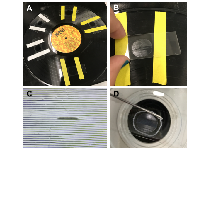
1. Place strips of tape 1.5-2 inches apart perpendicular to the grooves on the vinyl record’s grooved surface, as shown in Figure 1.A.
2. Pipette a drop of melted agarose onto the vinyl record between the strips of tape, and immediately add a microscope slide on top of the drop to make a pad against the vinyl record.
3. After cooling, remove microscope slide from the vinyl record by lifting from one side as shown in Figure 1.B. If the pad is too large, it can be trimmed down by using another microscope slide’s flat edge to make straight cuts.
4. Add a volume of 2-5l of anesthetic at a time, depending on the size of your pad. Then pick worms onto anesthetic droplet (the grooves are an optimal width for hermaphrodites from L4-adult).
5. Position worms into the grooves in the agarose pad utilizing the minutien pin (Figure 1.C). Worms are easier to position when most of the anesthetic has been absorbed into the pad, but don’t allow it to dry completely, as this will form air bubbles in the adjacent grooves.
6. Make a circle line around the pad with the syringe filled with Vaseline before placing the coverslip as shown in Figure 1D. Slightly press the coverslip over the pad and vaseline to keep the worms in place and make a seal around the pad to prevent evaporation of the anesthetics mix.
This protocol can be used in conjunction with microbeads, but in our hands they do not seem to improve the immobilization of the worms.
The vinyl record grooved pads function, in part, as a poor-person’s microfluidics for live imaging. It allows the placing of worms into single channels organized as in a microfluidic chip, thus reducing the time searching for the worms for imaging on the slide. Addition of drugs or food (for prolonged imaging) could potentially be done with a fine needle inserted through the Vaseline ring. An additional advantage of these pads is that there is no difference in fluorescence brightness loss compared to the conventional agarose pad.
This protocol’s affordability, the ease of use and training has simplified our live imaging of m+z- males expressing low fluorescence fusion proteins in the germline significantly.
We thank the Mikaela Murph and Erlyana Clarke for trying the protocol and providing us with feedback.
Figures
References
Zhang, M., Chung, S. H., Fang-Yen, C., Craig, C., Kerr, R. A., Suzuki, H., … Schafer, W. R. (2008). A self-regulating feed-forward circuit controlling C. elegans egg-laying behavior. Current Biology : CB, 18(19), 1445–1455. 