Worm Breeder's Gazette 9(2): 49
These abstracts should not be cited in bibliographies. Material contained herein should be treated as personal communication and should be cited as such only with the consent of the author.
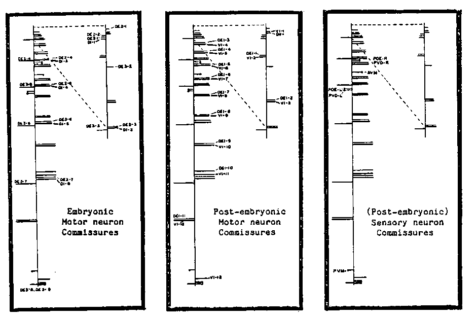
The nervous system of Ascaris contains 298 neurons each of which is
an identified cell whose morphological features are reproducible from
animal to animal.  For some years, we have been analyzing the pattern
of motor neuron commissures in Ascaris.  (Commissures are single
processes which exit from the ventral nerve cord and run around the
body, either to the right or to the left, to the dorsal cord).
Although in general the commissure of each motor neuron is
reproducibly to the right or to the left, we have found rare
individuals in which the handedness of specific commissures is
reversed.  These exceptions fall into two classes.  The first class
includes animals in which a single commissure or commissure pair is
reversed (Table 1).  Most of these involve the reversal of normally
righthanded VI and DE1 commissures to produce single lefthanded VI
commissures or lefthanded VI/DE1 commissure pairs.  Righthanded DE1
commissures are almost never observed to reverse alone, although 4
examples of normally lefthanded DE1 commissures being reflected to the
right have been observed.  Single reversals are seen most frequently
in VI/DE1 pairs adjacent to two lefthanded VI/DE1 commissure pairs
which occur normally in the head and in the tail (Table 2); perhaps
they reflect the spread of a local 'reversing' factor present at these
sites.  Selective reversal of righthanded VI commissures and
lefthanded DE1 commissures suggests that the factor's influence may be
stronger on VI than on DE1 motor neurons.
The second class consists of variant animals in which entire sets of
motor neurons have reversed commissures.  Initially, we found 2 Ab
stained preparations in which all DE2, DE3 and DI commissures were
reversed whereas all DE1 and VI commissures have their normal
handedness.  Subsequent screening of 1639 animals with darkfield
optics (which allows commissures to be viewed in live animals) has
revealed 2 additional animals with identical patterns of commissure
reversals as well as 6 other animals in which the commissures of all
motor neurons are reversed.  Since in C.  elegans, neurons analogous
to the DE2, DE3 and DI neurons are generated during embryogenesis
whereas the DE1 and VI neurons are from post-embryonic lineages, we
refer to these two types of variant animals as MIR-EMB and MIR-ALL
respectively.  Neither MIR-EMB nor MIR-ALL animals have any apparent
behavioral alteration.
Analysis of other neurons which have asymmetric shapes has shown
that two other embryonic neurons in the head (analogous to RID and
SABD) have mirror image morphologies in both MIR-EMB and MIR-ALL
animals and that cells analogous to the (postembryonic) AVF cells are
normal in MIREMB animals, but mirror images in MIR-ALL variants.
Neurons with asymmetric commissures in the tail are also inverted in
MIR-ALL animals (MIR-EMB animals have not yet been examined.  In
contrast to these alterations in the shapes of neurons, we find that
the location of asymmetrically positioned neuronal cell bodies (
Ascaris analogues of the PDEL/PDER, PVDL/PVDR, PVM/AVM and AQR/PQR
neurons) are not reversed in either of the variants.  (The nucleus of
the excretory duct, on the other hand, is on the left in normal and
MIR-EMB animals, but on the right in MIR-ALL animals.) Thus it appears
that MIR animals are not complete mirror images of the normal animals,
but rather that they are variants with mirror image neuron
morphologies.
The existence of two classes of MIR variants suggests that there are
two separate mechanisms for determining the handedness of neurons: one
operating during the growth of embryonic neurons, the other affecting
the post-embryonic neurons, possibly reflecting maternal vs. zygotic
expression of a common gene.  Confirmation of this possibility awaits
genetic experiments in Ascaris or the isolation of comparable variants
in C.  elegans.[See Figure 1]
[See Figure 2]
[See Figure 3]