Worm Breeder's Gazette 9(1): 62
These abstracts should not be cited in bibliographies. Material contained herein should be treated as personal communication and should be cited as such only with the consent of the author.
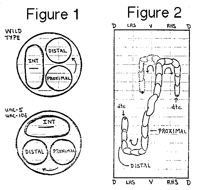
The products of the unc-5 and unc-106 genes are required for correct
dorsal/ventral positioning of longitudinal nerves and for dorsally-
directed, circumferential growth of motor axons from cell bodies in
the ventral hypodermis (1.2,3,4).  In addition, the unc-106 product is
required for ventrally-directed, circumferential growth of sensory
axons from cell bodies in the lateral hypodermis (5).  Certain non-
neuronal cells (head mesodermal cell and excretory cell) with
attachments to the body wall are also frequently displaced in unc-5
and unc-1O6 mutants.  All of the mutant defects might be explained if
the products of these genes mark dorsal/ventral position on the body
wall or help decode such information.  Recently, we observed that the
reflexions of the hermaphrodite and male gonads are abnormal in unc-5 (
e53) and unc-106 (ev400) mutants.  In normal development the anterior
and posterior arms of the hermaphrodite gonad are produced by active
migrations of the distal tip cells along the ventral body wall (6).
Late in the third larval stage these cells make dorsal = clockwise
turns (relative to the body wall, see Figure 2) to produce a mature
gonad with the distal (reflexed) arms positioned dorsal to the
proximal arms (Figure 1).  In unc-5 and unc-106 mutants, the
reflexions occur at the normal time.  For about one half of the distal
tip cells, the direction of turning is normal.  In the remaining cases,
however, the cells turn ventral = counterclockwise on the body wall
to produce a mature gonad with the distal arms positioned lateral to
the proximal arms (Figure 1).  Similar mistakes are made by the linker
cells in males of these mutants.  The distal tip cells in
hermaphrodites and the linker cell in males appear to be closely
apposed to the body wall during gonad extension and reflexion (as
judged by light microscopy).  The position of reflexion may be
determined by a timing mechanism as there is no apparent discontinuity
on the body wall.  The direction of reflexion, consistently dorsal =
clockwise in wild type, appears to be random in unc-5 and unc-106
mutants.  It is intriguing to speculate that the leading cells of the
gonad contact the body wall to obtain some of the same positional
information as used by the developing nervous system.  We hope soon to
examine these contacts by electron microscopy.