Worm Breeder's Gazette 8(3): 54
These abstracts should not be cited in bibliographies. Material contained herein should be treated as personal communication and should be cited as such only with the consent of the author.
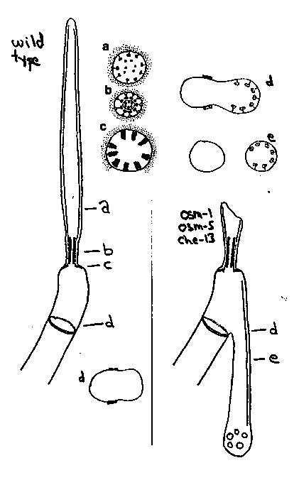
The amphid channel cilia are about 7.5 microns long.  Nine doublet-
microtubules extend for most of the length.  These microtubules are
attached to the ciliary membrane by Y-links (a).  Near the base of the
cilia, the doublet-microtubules are drawn together by a cylindrical
structure, called the apical ring (b).  The Y-links are pulled inward
and reveal their true shape.  A variable number of small singlet-
microtubules originate on the inner face of the ring.  Below the
apical ring the Y-links are elaborated into transitional fibers which
connect the doublet microtubules to the membrane (c).  There are no
basal bodies.  In three mutants che-10(e1805), 8),
and osm-5 (p813), all of the sensory cilia assemble abnormally.  The
phenotypes are similar for all three mutants and, for simplicity, we
describe the channel cilia only.  The proximal structures including
the transitional fibers and the apical ring are present and
essentially normal.  The central microtubules end with the ring
however, and the outer doublets extend only a little further.
Surprisingly, the bulk of the missing microtubules can be accounted
for by doublet-microtubules than assemble ectopically behind the cilia
(d).  These doublets are attached to the membrane by Y-links and
allign longitudinally to make a cilia-like projection directed
posteriorly in the sheath cell (e).  Like the normal cilia, these
ectopic projections are topologically distal to the neuron/sheath
junctions.  We suggest that the microtubules and their links self-
assemble and the wild type che-10,
are needed to ensure that they assemble only on the correct template.

[See Figure 1]