Worm Breeder's Gazette 7(2): 38
These abstracts should not be cited in bibliographies. Material contained herein should be treated as personal communication and should be cited as such only with the consent of the author.
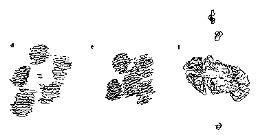
The meiotic chromosomes of C.  elegans males were reconstructed from
electron micrographs of serial sections of three primary spermatocytes.
Viewed from the spindle pole the chromosomes form a distinctive
pattern on the metaphase I plate.  The five autosomal bivalents form a
ring that surrounds the centrally placed X univalent.  Figure a is a
computer reconstruction of the five autosomes and fig.  b is a display
of four of the autosomes and the X.  The autosomal half-bivalents lie
to either side of the equatorial plate (as shown for two autosomes in
fig.  c.) and the X lies on the equatorial plate.  The metaphase II
spindle in each lobe of a bilobed secondary spermatocyte was also
reconstructed.  One spindle contained six and the other five pairs of
chromosomes, the X had not disjoined at anaphase I.  When six
chromosomes are present five chromosomes form a ring around the sixth
chromosome (figs.  d and e).  The X cannot be distinguished from the
autosomes at this time.  Figure f.  is a side view of the metaphase II
spindle.
Kinetochores could not be distinguished on these chromosomes,
prepared by conventional electron microscopic techniques.
Microtubules appeared to insert directly into the chromosome along the
poleward face.
Centrioles were present in the asters of the meiotic spindles in
primary and secondary spermatocytes of both males and hermaphrodites.
The overall shape of the spindles was similar to the mitotic spindles
of C.  elegans.  In contrast, the meiotic spindles seen in oocytes
have broad ends and are barrel-shaped similar to the meiotic spindles
of mammalian oocytes.  Centrioles are absent from the poles of barrel-
shaped spindles in mammalian oocytes, and have not been seen in the
barrel-shaped spindles of C.  elegans.  The time at which centrioles
are lost in the hermaphrodite gonad has not been determined precisely.
Centrioles are seen adjacent to pachytene nuclei in L4 hermaphrodites
during spermatogenesis, but have not been seen in young adult
hermaphrodites completing the spermatogonial divisions and beginning
oogenesis, or in older adult hermaphrodites.
[See Figure 1]
[See Figure 2]