Worm Breeder's Gazette 7(1): 89
These abstracts should not be cited in bibliographies. Material contained herein should be treated as personal communication and should be cited as such only with the consent of the author.
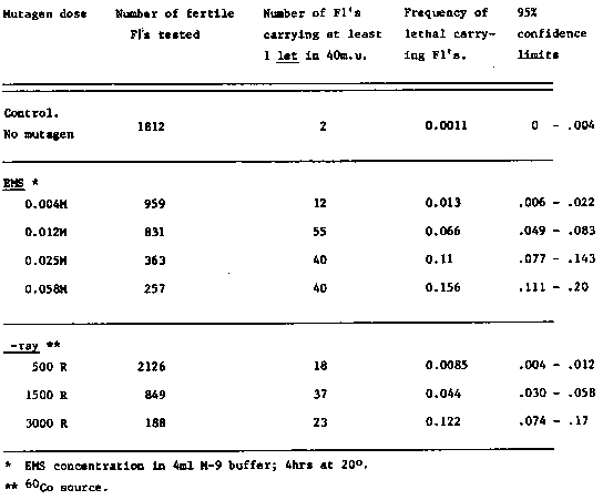
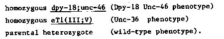
In order to obtain EMS and gamma-ray dose-response curves for the
induction of mutations in C.  elegans, we are measuring the frequency
of recessive lethals induced within a defined region of 40 map units.
Using the translocation, eT1(III;V), as a balancer (Rosenbluth and
Baillie, CSH meeting 1981), we simultaneously screen the right half of
LGIII and the left half of LGV for lethals as follows.
Due to the segregation patterns of eT1 heterozygotes, and due to the
fact that as a heterozygote eT1 suppresses crossingover between sma-2
and unc-64 on LGIII and between unc-60 and dpy-11 on LGV, heterozygous
hermaphrodites of the genotype, dpy-18/eT1(III);unc-46/eT1(V) produce
only three types of self
offspring:
[See Figure 1]
Consequently, if such a heterozygote carries a lethal mutation on
either the dpy-18 or unc-46 marked chromosome,- anywhere in the
crossover suppressed region, - there will be no Dpy-18
Our current protocol consists of treating adult heterozygous
hermaphrodites (of the above genotype) with a given dose of mutagen
and allowing these to 'self' at 20 C.  Single F1 heterozygotes (wild-
type) are then placed on individual plates and their progeny are
screened for the absence of DpyUnc individuals.  Strains from F1's
producing no DpyUnc's are putatively lethal bearing and are maintained
by repicking heterozygotes.  If no DpyUnc's appear among at least 250
offspring, the F1 is considered to have carried at least one lethal
mutation somewhere within the crossover suppressed regions (or within
1m.u. Of them.)
The data collected so far are summarized in the following table.
[See Figure 2]
It should be noted that with the current protocol, half the
chromosomes screened were mutatgenized in sperm and half in oocytes.
Preliminary experiments by others in our laboratory (Rogalski and
Pilgrim) indicate that the oocytes are much less sensitive to gamma-
radiation than are the sperm ( as is the case in Drosophila).  We are
planning to measure the frequencies in sperm and oocytes separately.
If the above values for the gamma- radiation are only due to lethals
induced in sperm, then they are only half the true sperm lethal
frequency.
We hope that this type of data will provide some guide lines for the
choice of EMS and gamma-ray doses in future experiments.