Worm Breeder's Gazette 2(2): 30
These abstracts should not be cited in bibliographies. Material contained herein should be treated as personal communication and should be cited as such only with the consent of the author.
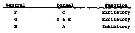
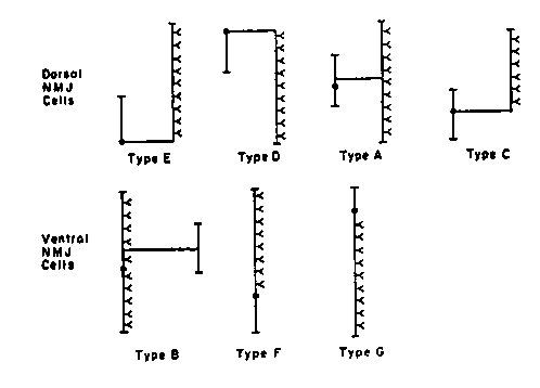
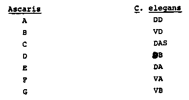
The motor nervous system of Ascaris consists of five sets of
segmented neurons each containing eleven cells that make synapses onto
muscle, together with six ventral interneurons that make synapses onto
some of the motorneurons.  The neurons can be divided into seven
classes (see figure) that appear to be structurally identical to the
seven classes in C.  elegans, with the following equivalences .
[See Figure 1]
The dorsal n.m.j. cell types C, D and E receive interneuron input,
but the A cell receives synapses only from ventral motorneurons.  The
ventral F and G cells receive input from interneurons; the B cells
receive synapses only from dorsal motorneurons.  All these output
cells make synapses with muscle only in one nerve cord.  Each segment
has one copy of cell types A, D and E, and two copies of types B, C, F
and G.
Anatomical similarities (relative positions in nerve cords; details
of the patterns of neuromuscular connectivity) are used to assign
functional equivalences in the dorsal and ventral cords.
[See Figure 2]
The function of types of A, B, C, D and E neurons has been
determined physiologically (see Walrond and Kass).
The motorneuron synapses onto the inhibitory neurons provide
pathways for reciprocal inhibition, between both cords, that is
generated peripherally rather than centrally.
[See Figure 3]