Worm Breeder's Gazette 12(4): 70 (October 1, 1992)
These abstracts should not be cited in bibliographies. Material contained herein should be treated as personal communication and should be cited as such only with the consent of the author.
The gene pat-3 (aka epi-2 )encodes a ß1-like integrin subunit (S. Gettner, pers. comm.). Three mutant alleles have been isolated. The strongest allele, pat-3 ( rh54 ),is embryonic lethal, and affects development of most muscles, plus cell migrations and attachments in non-muscle tissues. pat-3 ( rh151 )is viable but sterile, and does not appear to disrupt body muscle cells. The mildest allele, pat-3 ( rh96 ),is viable. Using serial thin section reconstruction of adult genetic mosaics by electron microscopy, we compared MSpp- and MSpa- mosaics to determine the role(s) of ß-integrin in muscle development, and AB p1 -mosaics to examine alterations in excretory canal cell development.
Normal body muscles adhere to the body wall cuticle via fibrous linkages to membrane densities along the inner surface of the muscle's plasma membrane. A normal cell contains several sarcomeres lying oblique to the body axis, also anchored to membrane densities. The rh54 mutant phenotype is cell autonomous. Affected cells are not contractile. They detach from the body wall, but remain in close contact with nearby wild type muscle cells, forming both gap junctions and adherens junctions with them and with each other. Mutant cells are longer and much thinner than normal. No sarcomeres are formed, but separate bundles of thick filaments, thin filaments, and dense aggregates are found in the cytoplasm. Some cells form branches which extend dorsoventrally to the opposite muscle quadrant. Interestingly, mutant cells still form "muscle arms" which extend in normal fashion to the nerves and nerve ring in order to receive synaptic inputs. The lethal myospheroid ,ß-integrin mutant in Drosophila causes similar defects in muscle development (Volk et al., Cell 63:525-536, 1 990).
The normal excretory canal cell is H-shaped, with its soma lying just behind the nerve ring in a ventral position. Four tube-like canals emerge laterally from the soma and join the lateral hypodermis before turning to run towards the nose or tail within the hypodermal cords. Each canal is distinguished by a single lumen which appears to condense from a network of many short membrane bound tubules. The contents of these four excretory cell canal lumens are dumped jointly via the excretory pore to the exterior of the animal. The function of the excretory system is uncertain, but may represent the equivalent of a nematode kidney. In AB p1 -mosaics, the excretory cell does not extend canals away from the soma. Instead the soma is greatly enlarged, with several multiply branched lumens winding within the soma. A normal connection from these lumens to the excretory pore is still achieved. The plasma membrane of the soma is often heavily invaginated, as if the cell had produced excess surface area in anticipation of extending canals. The total cell volume of the mutant canal cell appears to be equivalent to wild type canal cells.
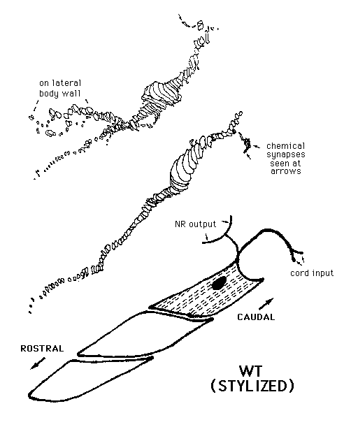
Several other organ systems are also disrupted cell autonomously by mutation. Sex muscles fail to spread out properly, the anal depressor muscle often loses anchorage, and the ovarian sheath cells don't mange to encircle the gonad properly - hence the epi-2 phenotype. The distal tip cell is still motile, but fails to constrict the tip of the gonad very well, leading to an abnormally large gonadal cylinder. [See Figure]
Volk et al., Cell 63:525-536, 1 990.