Worm Breeder's Gazette 11(4): 81
These abstracts should not be cited in bibliographies. Material contained herein should be treated as personal communication and should be cited as such only with the consent of the author.
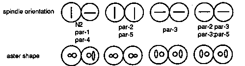
In an effort to understand the relationships among the par genes we
have been analyzing double mutant combinations.  The phenotypes of
mutants in the five par genes are similar but readily distinguishable
from one another.  Strong loss-of-function mutations in all genes
affect cleavage spindle position and orientation, cleavage rate, P
granule localization, and development of the intestine but do so in a
gene-specific manner.  We have interpreted these phenotypes to
indicate that the par genes encode components of a maternally provided
system for establishing cleavage asymmetry and cytoplasmic
localization in the early embryo.
The results from the five doubles we have examined thus far indicate
that the genes are not in a simple dependent pathway but they do
interact.  There are no clear epistatic relationships, although
individual aspects of the phenotypes may show epistasis (see below);
the cellular phenotypes of the strong mutants are simply additive, not
synergistic.  However, we often see synergism with respect to
viability in double mutant combinations of weak alleles.  The most
striking example is the double homozygote par-2(it5ts);
7ts).  it5 gives greater than 70% viable progeny at
15 C and it57 gives nearly 100% viable progeny at 15 C.  it5;it57
doubles give almost complete lethality at 15 C.  We also see dominant
enhancement of weak alleles of one par by one copy of a strong allele
at a second par gene.
Two particularly interesting double mutant combinations are par-2
par-3 and par-3;par-5.  par-3 affects the orientation of the second
cleavage spindles in a way that is opposite to par-2 and par-5.[See
Figure 1]
We were surprised to find that par-3 is epistatic to par-2 and par-5
for this particular phenotype.  This indicates that none of the three
genes are required for the rotation of the P1 spindle.  Rather, par-3+
acts to prevent spindle rotation and par-2+(5+) acts to insure that
par-3+ acts only in the AB.  This could result either from
localization of par-3+ activity to the AB or by action of par-2+(5+)
in the P1.
We think it likely that par-2+(5+) acts in the P1 to inhibit or
modify the action of par-3+, because we have obtained evidence that
par-3+ activity is present in P1.  In the wild type, P1 spindle
rotation occurs reproducibly in embryos that are slightly flattened (
squashed) by pressure from the coverslip.  Our initial observations of
par-3 mutants were done on unsquashed embryos.  When we observe par-3
mutant embryos under squashed conditions, the orientations of AB and
P1 spindles are much more variable than under unsquashed conditions.
We conclude, therefore, that although the rotation of the P1 spindle
does not absolutely require par-3+ the strength of the rotation or the
stability of the new spindle position is dependent upon par-3+
function.  Perhaps par-3+ encodes an element of the cytoskeleton that
normally stabilizes spindle position in the early embryo.  par-2+ then
acts in the P1 to modify the par-3+ activity allowing the spindle to
rotate.  Another aspect of the par phenotype may relate to this point.
In the wild type, the asters of the first cleavage spindle show
different behavior in P1 and AB.  In the P1 cell the aster becomes
disc shaped while in the AB the aster remains spherical.  In par-1 and
par-4, the P1 aster becomes disc shaped.  In par-3, both AB and P1
asters become disc shaped, although not to the same extent as in wild
type.  In par-2 and par-5 both asters remain spherical.  In the par-2
he asters are disc shaped (par-3;par-5 has
not yet been checked).  Thus the change in aster shape at the end of
the first division seems to correlate with spindle rotation.