Worm Breeder's Gazette 11(4): 8
These abstracts should not be cited in bibliographies. Material contained herein should be treated as personal communication and should be cited as such only with the consent of the author.
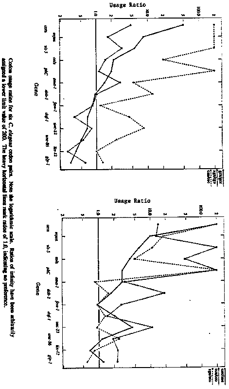
A general trend from highly asymmetric codon usage in strongly
expressed genes to less asymmetric codon usage in weakly expressed
genes is observed in many organisms (P.  Sharp et al., Nucl.  Acids
Res.  17 (1988) 8207-8211).  Sequence data are now available for worm
genes representing a number of gene families, with a variety of
expression patterns; hence one can look for variations from the
distinctively biased 'average' C.  elegans codon usage pattern (WBG
Jan.  1990).
Large differences in codon usage are not apparent between members of
the myosin, collagen, actin, and lin-12-glp-1 gene families; however,
there are some striking differences between gene families.  Some of
these are summarized in the figures on the following page, which show
usage ratios for pairs of Ala, Arg, Leu, Phe, Pro, and Ser codons.  In
every case, the usage asymmetry is generally higher in the actin,
myosin, and collagen families, and lower in unc-86 and the lin-12-glp-
1 family.
The usage ratios for the Ala and Arg pairs shown (gca/gcc and
aga/cgu) actually reverse, indicating a shift in codon preference.
The most significant shift is in the Arg codons of daf-1,lin-12, and
glp-1, in which terminal purines are preferred over terminal
pyrimidines.  The preference for terminal a's in Gly and Pro codons,
however, is decreased in these genes relative to that found in the
actins and collagens, so the shift is not a general one towards third-
position purines.
These data - especially the strong reversals in preference - suggest
that codon usage asymmetry, at least for some amino acids, may play a
role in regulating gene expression.  They also make it fairly clear
that codon usage asymmetry will be a poor guide to the identification
of coding exons in some genes.  It will be interesting to see whether
other ways of looking at codon data reveal additional patterns.
Sources of the data are as follows: act-1, act-2, act-3, act-4 (Krause
etet a al., J.  Mol.  Biol.  208 (1989) 381-392); ama-1 (Bird and Riddle,
Mol.  Cell.  Biol.  9 (1989) 4119-4130); col-1, col-2 (Kramer et al.,
Cell 30 (1982) 599-606); col-6, col-7, col-8, col-14, col-19 (Cox et
al., Gene 76 (1989) 331-344); daf-1 (Georgi et al., Cell 61 (1990) 635-
645); deb-1 (Barstead and Waterston, J.  Biol.  Chem.  264 (1989)
10177-10185); dpy-13 (von Mende et al., Cell 55 (1988) 567-576); fem-1 (
Spence et al., Cell 60 (1990) 981-990); glp-1 (Yochem and Greenwald,
Cell 58 (1989) 553-563); lin-12 (Yochem et al., Nature 335 (1988) 547-
550); myo-1, myo-3 (Dibb et al., J.  Mol.  Biol.
205 (1989) 603-613); pkC (Gross et al., J.  Biol.  Chem.  265 (1990)
6896-6907); sqt-1 (Kramer et al., Cell 55 (1988) 555-565); unc-15 (
Kagawa et al., J.  Mol.  Biol.  207 (1989) 311-333); unc-22 (Benian et
al., Nature 342 (1989) 45-50 and this WBG); unc-54 (Karn et al., Proc.
Natl.  Acad.  Sci.  USA 80 (1983) 42534257); unc-86 (Finney et al.,
Cell 55 (1988) 757-769); and vit-5 (Spieth et al., Nucl.  Acids Res.
13 (1985) 7129-7138).
[See Figure 1]