Worm Breeder's Gazette 11(4): 10
These abstracts should not be cited in bibliographies. Material contained herein should be treated as personal communication and should be cited as such only with the consent of the author.
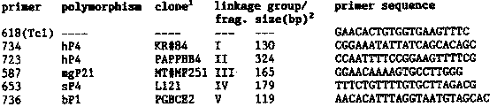
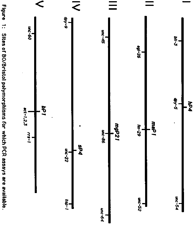
After mapping many lethal mutations by standard methods, we decided
to try another approach to genetic mapping which has worked well and
may be of general interest. Polymerase chain reaction (PCR) assays are
being developed to specifically detect several Bristol/Bergerac Tc1
polymorphisms on each of the 5 autosomes (Fig.1). The segregation of
these polymorphisms can be followed in the progeny of Bristol/Bergerac
hybrids by doing PCR reactions with DNA from single worms. Each assay
consists of a unique primer flanking the Tc1 insertion site and a
common primer complementary to internal Tc1 sequence. The Bergerac
allele is detected by amplification of a DNA fragment of
characteristic size; no amplification occurs from the Bristol allele.
For more efficient mapping, flanking primers were chosen so that
amplification products from different polymorphisms are easily
distinguishable by size on a non-denaturing acrylamide minigel. This
allows several assays to be used in the same PCR reaction. To map a
mutation, heterozygous Bristol males are crossed into the Bergerac
strain RW7000, and an F1 hermaphrodite that segregates mutants is
isolated. 20 - 30 of F2 mutant homozygotes are picked (dead or alive!)
to separate tubes and their DNA is subjected to PCR analysis with the
assay set of primers (see below). For unlinked polymorphisms, the
Bergerac allele is detected in 75% of the progeny. This frequency is
reduced for linked polymorphisms, and approaches 0% in cases of tight
linkage. Additional primer sets are being developed to provide 4 - 5
markers per chromosome. For these we are using BO clones containing
Tc1 elements and determining their physical map position to identify
well placed polymorphisms. For those interested in trying this method
we intend to make a starter kit containing each of the six primers
below.
reference clone in the physical map data base. We would like to
thank A.Rose, M.Papp, M.Finney, D.Baillie and M.Krause for providing
us with their clones containing the polymorphic sites.
size of amplified fragment when primer is used with Tc1 primer 618.
PCR reaction conditions: 2.5  l of worm lysis buffer containing a
worm, 25 picomoles of each primer, 2.5  l 1OX standard PCR reaction
buffer (100 mM Tris pH 8.3, 500 mM KCl, 15 mM MgC12, 0.01%[w/v]
gelatin), 4  l dNTP mix (1.25 mM for each dNTP), 0.625 units of Taq
polymerase, H20 to 25  l. 30 cycles: 94 C, 30 sec; 58 C, 60 sec; 72 C,
60 sec.
Single worm PCR (from R. Barstead): Worm is picked into worm lysis
buffer (50 mM KCl, 10 mM Tris pH 8.3, 2.5 mM MgC12, 0.45% NP-40, 0.45%
Tween 20, 0.01% gelatin, 60  g/ml proteinase K), frozen at -70 C for
10 min, overlaid with a drop of oil, heated to 60 C 1 hr and then 95 C
for 15 min.