Worm Breeder's Gazette 10(1): 89
These abstracts should not be cited in bibliographies. Material contained herein should be treated as personal communication and should be cited as such only with the consent of the author.
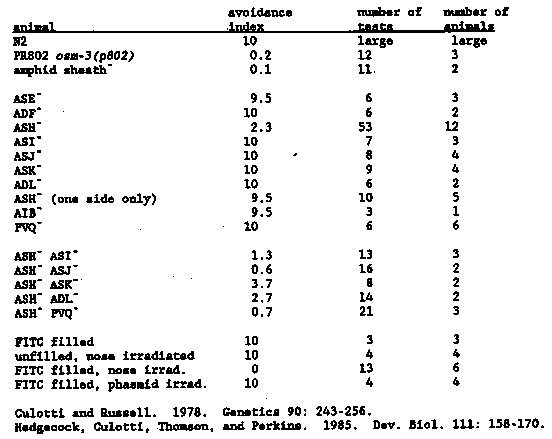
When a worm encounters a region of high osmotic strength with the
tip of its nose, it responds by backing up, typically several body
lengths, and usually turning around.  Genetic analysis of this
behavior should be powerful, since selection of animals that fail to
respond to osmotic strength is simple.  To determine which sensory
neurons mediate the osmotic avoidance response (hereafter called the
Osm phenotype), we used a laser to kill neurons in early L1 larvae,
and performed single-animal Osm assays on the animals as adults.  For
a discussion of various concerns about the validity of laser-killing
experiments on neurons, see the accompanying newsletter entry (Thomas,
Garriga, Avery, and Horvitz).
Our assay is a modification of that used by Culotti and Russell (
1978).  About 15 l of 4 M fructose (plus Congo Red, a dye that worms
do not respond to) is applied to a dry NG plate in a ring with an
inner diameter of about 0.8 cm and allowed to soak into the agar.  A
single animal is moved without bacteria onto the test plate inside the
osmotic ring and allowed to run for 15 minutes, with visual checks at
least once per minute (tracks are observed at the end of the assay to
be sure that a transient crossing did not occur).  The time the animal
takes to cross the osmotic barrier is recorded, rounded to the nearest
minute.  The minimum score is one minute.  Notes on individual
encounters are usually made as well.  A wild-type animal usually
encounters the fructose ring in fewer than 30 seconds, responds
normally, and heads off in a new direction.  In a typical assay the
animal will encounter the ring about 20 times in 15 minutes.  Wild-
type animals very rarely cross the ring, and then only very late in
the assay (when the osmotic barrier has diffused considerably).  When
a mutant such as osm-3(p802) is run in this assay, the animal
typically crosses the barrier in less than one minute, without any
response [See Figure 1].  From the results of several such tests we
calculate an osmotic avoidance index (OAI) - 10/14 (t - 1), where t is
the mean time to cross.  This produces a range of OAI from 0 to 10.
The OAI for N2 is very close to 10 and that of osm-3(p802) is 0.2 (osm-
3 animals never respond to the osmotic barrier, but sometimes take
longer than one minute to cross).
What sensory neurons mediate this behavior?  We knew the sensing was
likely to be in the head, and the only known head defect of osm-3
animals is ultrastructural abnormalities in the amphid sensory endings.
Therefore we killed the amphid sheath cell (both sides) to test
whether the Osm response was eliminated.  It was (see Table for a
summary of all of the cell killing data).  Since the sheath acts as a
glial-like sheet around all eight of the amphid chemosensory endings,
it seemed likely that one or more of these neurons was mediating Osm.
We FITC-filled six of the amphid chemosensory neurons (ADF, ASH, ASI,
ASJ, ASK, ADL), by the method of Hedgecock et al.  (1985), and
bleached the dye using a fluorescein filter set and an epifluorescence
light source.  This treatment also eliminated the Osm response (see
Table for data and controls).  With the laser we killed seven of the
eight amphid chemosensory neuron types individually and tested each
for osmotic avoidance (the eighth neuron, ASG, has not been done
because it is more difficult to identify with certainty and is not
FITC-filled.) In addition, the amphid interneurons AIB and PVQ were
killed.  Only when ASH was killed were animals significantly less
responsive than normal.  ASH animals frequently failed to respond to
the osmotic barrier at all, but sometimes responded and crossed only
later in the assay.  Even when an ASH animal responded it was usually
deeper in the osmotic barrier than normal, suggesting a less sensitive
or slower response.  Killing ASH reduces the efficacy of the Osm
response, but it is not the whole story.
Any other sensory neuron involved in the Osm response should also be
in the amphid, and probably one of the FITC-filled cells, because of
the amphid sheath and FITC-filling results.  We searched for a second
neuron by doing double kills with ASH (both sides) and a second amphid
sensory neuron (both sides).  Of the four neurons tested, only killing
ASJ effectively eliminated the residual Osm response (we are not yet
sure whether the apparent reduction by killing ASI is significant; we
have not yet tested ADF).  ASJ has output only to PVQ, an interneuron.
Killing PVQ and ASH also eliminated the residual Osm sensitivity,
suggesting that ASJ acts through PVQ.  We should emphasize that the
assignment of ASJ and PVQ to the Osm response is tentative; we must
kill these cells together with ASH in more animals to be certain of
this relatively marginal effect.
We anticipate that further application of the laser to the Osm
behavior will clarify the rest of the neuronal pathway from sensory
input to motor response.  Identification of the cells in this pathway
will assist in the interpretation of mutants defective in the Osm
response.