2 Department of Biochemistry and School of Biological Sciences University of Utah, Salt Lake City, UT, USA
Transmission electron microscopy (TEM) is used extensively to study the nematode C. elegans. A typical specimen preparation for TEM involves chemical fixing followed by consecutive changes of reagents and embedding in plastic before thin sectioning. To handle this extremely small animal (~ 1 mm in length and 50 μm in diameter for adults), the most popular method is to immerse chemically fixed worms in warm agarose (Hall et al., 2012). Upon cooling, the solidified block is then trimmed to a maneuverable size. The goal and merit of this method is to avoid losing precious specimens during solution change and specimen transfer, as well as to facilitate desired body orientation prior to sectioning. Here we report the use of hollow fibers in lieu of agarose to achieve the same goal.
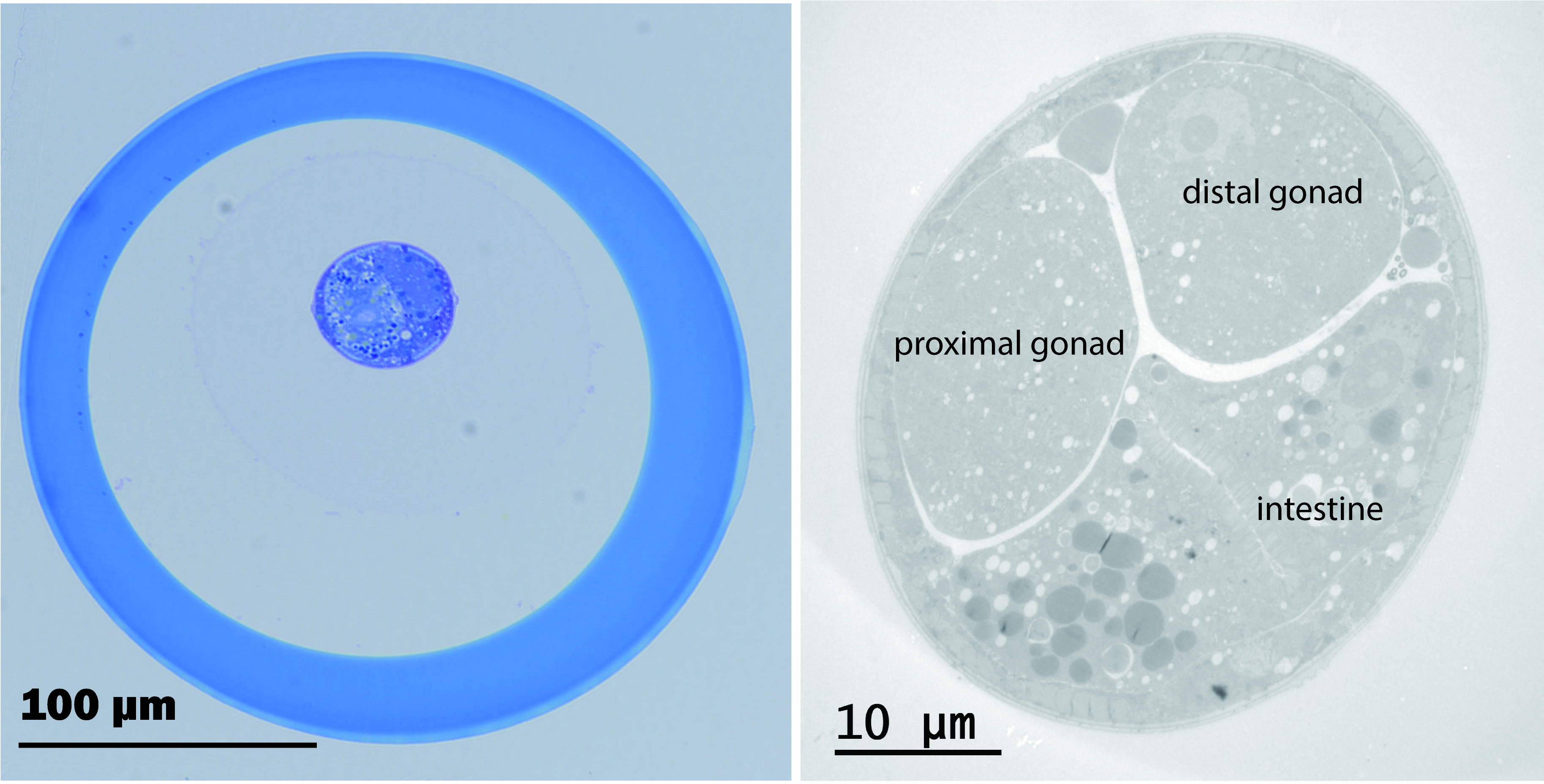
We used transparent hollow fibers from a hemodialyzer (Figure 1A) to encapsulate the worm. Each fiber has an inner diameter of 175 μm and wall thickness of 25 μm. For easy manipulation of fibers, an insect pin1 of 100 μm in diameter is inserted into the lumen of the fiber (Figure 1B, Video 1). To encapsulate, a piece of appropriately fixed worm specimen is transferred to a ~ 3μl drop of buffer rinse and then one end of the hollow fiber is submerged into the drop. With capillary action, the specimen is drawn up into the fiber (Video 1). This loaded fiber is handled with tweezers and processed normally as for tissue samples2. At the end of resin infiltration, excess length of the fiber is trimmed off and the worm can be oriented as desired in a horizontal mold (Figure 1C). During curing, the specimen will sink into the bottom of the mold due to gravity. The fiber gives sufficient leeway (at least 25 μm) at the bottom of the block for trimming (Figure 1D). Therefore, pre-filling the mold or re-embedding the specimen is not necessary (Mulcahy et al., 2018, Muller-Reichert et al., 2003). This greatly expedites the thin-sectioning process. Figure 2A shows that the hollow fiber, made of ethylene vinyl alcohol copolymer, remains intact after exposure to osmium, uranyl acetate, alcohol, acetone and resin Embed 812. Fine TEM images of the worm (Figure 2B) indicate that the hollow fiber, with its molecular weight cutoff at ~30 kDa, allows for penetration of these chemicals. We have also successfully encapsulated the worm specimens by simple capillary action while they were soaked in 1:1 mixture of resin and acetone. Therefore, encapsulation is applicable to samples that are prepared by other means, for example by high pressure freezing and freeze-substitution with organic solvents.
1 A Minutiens insect pin is used (size 0.10, Austerilitz Insect Pins®). Individual pins are about 12 mm in length, 0.1 mm in diameter at the shaft, and 0.0125 mm in diameter at the tip (Figure 1). For easy manipulation of the pin, one end is heat annealed to a p10 pipette tip (Video 1).
2 We use ~2.5 cm length of the hollow fiber for encapsulation and place the loaded fiber into 2 ml microfuge tube for solution changes. If the worm is situated in the middle of the fiber, there is no worry of losing the specimen during processing. However, if the hollow fiber were to be trimmed shorter, both ends need to be sealed either by squeezing with forceps (Video 1) or by heating with hot platinum wire to prevent escape of the specimen.

References
Hall DH, Hartwieg E, and Nguyen KC (2012). Modern electron microscopy methods for C. elegans. Methods Cell Biol. 107, 93-149.

Mulcahy B, Witvliet D,Holmyard D,Mitchell J,Chisholm AD,Meirovitch Y, Samuel ADT, and Zhen M (2018). A pipeline for volume electron microscopy of the Caenorhabditis elegans nervous system.
Müller-Reichert T, Hohenberg H, O'Toole E T and McDonald K. (2003). Cryoimmobilization and three-dimensional visualization of C. elegans ultrastructure. J. Microsc. 212(Pt 1), 71-80.
Articles submitted to the Worm Breeder's Gazette should not be cited in bibliographies. Material contained here should be treated as personal communication and cited as such only with the consent of the author.
Leave a Reply
You must be logged in to post a comment.